Search your article
Peripheral Nervous System

Peripheral Nervous System
The peripheral nervous system (PNS) resides or extends outside the central nervous system (CNS), which consists of the brain and spinal cord. The main function of the PNS is to connect the CNS to the limbs and organs. Unlike the central nervous system, the PNS is not protected by bone or by the blood-brain barrier, leaving it exposed to toxins and mechanical injuries. The peripheral nervous system is divided into the somatic nervous system and the autonomic nervous system. [1]
In the somatic nervous system, the cranial nerves are part of the PNS with the exception of the optic nerve (cranial nerve II), along with the retina. The second cranial nerve is not a true peripheral nerve but a tract of the diencephalon.[2] Cranial nerve ganglia originated in the CNS. However, the remaining ten cranial nerve axons extend beyond the brain and are therefore considered part of the PNS.[3] The autonomic nervous system exerts involuntary control over smooth muscle and glands.[citation needed] The connection between CNS and organs allows the system to be in two different functional states: sympathetic and parasympathetic.
Somatic nervous system
The somatic nervous system includes the sensory nervous system and the somatosensory system and consists of sensory nerves and somatic nerves, and many nerves which hold both functions.
In the head and neck, cranial nerves carry somatosensory data. There are twelve cranial nerves, ten of which originate from the brainstem, and mainly control the functions of the anatomic structures of the head with some exceptions. One unique cranial nerve is the vagus nerve, which receives sensory information from organs in the thorax and abdomen. The accessory nerve is responsible for innervating the sternocleidomastoid and trapezius muscles, neither of which being exclusively in the head.
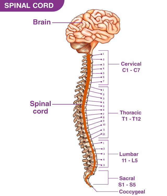
For the rest of the body, spinal nerves are responsible for somatosensory information. These arise from the spinal cord. Usually, these arise as a web (“plexus”) of interconnected nerves roots that arrange to form single nerves. These nerves control the functions of the rest of the body. In humans, there are 31 pairs of spinal nerves: 8 cervical, 12 thoracics, 5 lumbar, 5 sacral, and 1 coccygeal. These nerve roots are named according to the spinal vertebrata which they are adjacent to. In the cervical region, the spinal nerve roots come out above the corresponding vertebrae (i.e., nerve root between the skull and 1st cervical vertebrae is called spinal nerve C1). From the thoracic region to the coccygeal region, the spinal nerve roots come out below the corresponding vertebrae. It is important to note that this method creates a problem when naming the spinal nerve root between C7 and T1 (so it is called spinal nerve root C8). In the lumbar and sacral region, the spinal nerve roots travel within the dural sac and they travel below the level of L2 as the cauda equina.
Cervical spinal nerves (C1–C4)
The first 4 cervical spinal nerves, C1 through C4, split and recombine to produce a variety of nerves that serve the neck and back of head.
Spinal nerve C1 is called the suboccipital nerve, which provides motor innervation to muscles at the base of the skull. C2 and C3 form many of the nerves of the neck, providing both sensory and motor control. These include the greater occipital nerve, which provides sensation to the back of the head, the lesser occipital nerve, which provides sensation to the area behind the ears, the greater auricular nerve and the lesser auricular nerve.
The phrenic nerve is a nerve essential for our survival which arises from nerve roots C3, C4 and C5. It supplies the thoracic diaphragm, enabling breathing. If the spinal cord is transected above C3, then spontaneous breathing is not possible.
Brachial plexus (C5–T1)
The last four cervical spinal nerves, C5 through C8, and the first thoracic spinal nerve, T1, combine to form the brachial plexus, or plexus brachialis, a tangled array of nerves, splitting, combining and recombining, to form the nerves that subserve the upper-limb and upper back. Although the brachial plexus may appear tangled, it is highly organized and predictable, with little variation between people.
Lumbosacral plexus (L1–Co1)
The anterior divisions of the lumbar nerves, sacral nerves, and coccygeal nerve form the lumbosacral plexus, the first lumbar nerve being frequently joined by a branch from the twelfth thoracic. For descriptive purposes this plexus is usually divided into three parts:
- lumbar plexus
- sacral plexus
- pudendal plexus
Autonomic nervous system
The autonomic nervous system (ANS), formerly the vegetative nervous system, is a division of the peripheral nervous system that supplies smooth muscle and glands, and thus influences the function of internal organs. The autonomic nervous system is a control system that acts largely unconsciously and regulates bodily functions, such as the heart rate, digestion, respiratory rate, pupillary response, urination, and sexual arousal.[4] This system is the primary mechanism in control of the fight-or-flight response.
The autonomic nervous system is regulated by integrated reflexes through the brainstem to the spinal cord and organs. Autonomic functions include control of respiration, cardiac regulation (the cardiac control center), vasomotor activity (the vasomotor center), and certain reflex actions such as coughing, sneezing, swallowing and vomiting. Those are then subdivided into other areas and are also linked to autonomic subsystems and the peripheral nervous system. The hypothalamus, just above the brain stem, acts as an integrator for autonomic functions, receiving autonomic regulatory input from the limbic system.[5]
The autonomic nervous system has three branches: the sympathetic nervous system, the parasympathetic nervous system and the enteric nervous system.[6][7] Some textbooks do not include the enteric nervous system as part of this system.[8] The sympathetic nervous system is often considered the “fight or flight” system, while the parasympathetic nervous system is often considered the “rest and digest” or “feed and breed” system. In many cases, both of these systems have “opposite” actions where one system activates a physiological response and the other inhibits it. An older simplification of the sympathetic and parasympathetic nervous systems as “excitatory” and “inhibitory” was overturned due to the many exceptions found. A more modern characterization is that the sympathetic nervous system is a “quick response mobilizing system” and the parasympathetic is a “more slowly activated dampening system”, but even this has exceptions, such as in sexual arousal and orgasm, wherein both play a role.[7]
There are inhibitory and excitatory synapses between neurons. A third subsystem of neurons have been named as non-noradrenergic, non-cholinergic transmitters (because they use nitric oxide as a neurotransmitter) and are integral in autonomic function, in particular in the gut and the lungs.[9]
Although the ANS is also known as the visceral nervous system, the ANS is only connected with the motor side.[10] Most autonomous functions are involuntary but they can often work in conjunction with the somatic nervous system which provides voluntary control.